Laminitis Radiograph Measurements
Chronic Laminitis The Laminitis Site
Https Aaep Org Sites Default Files Issues 524 530 Pdf
Equine Medicine 2 Exam 3 Laminitis Ii Laminitis Ii Flashcards Memorang

Radiographic And Radiological Assessment Of Laminitis Sherlock 2013 Equine Veterinary Education Wiley Online Library
Chronic Laminitis The Laminitis Site
Herbie The Laminitis Site
This article will summarise the assessment of these 3 projections.
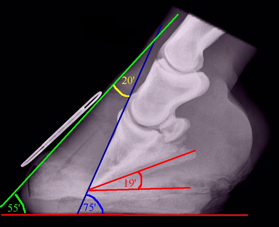
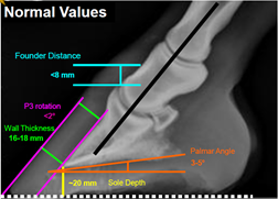
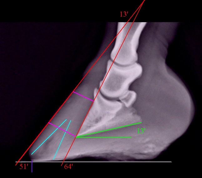
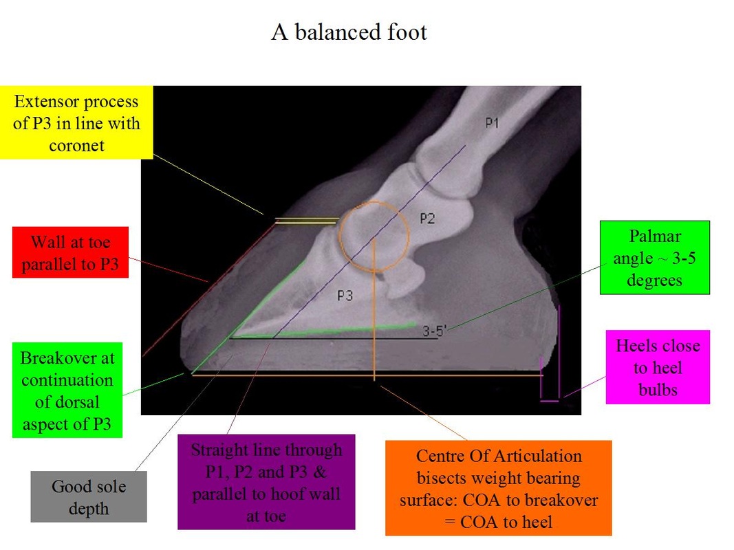
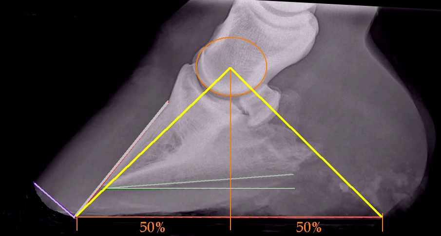
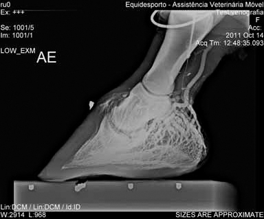
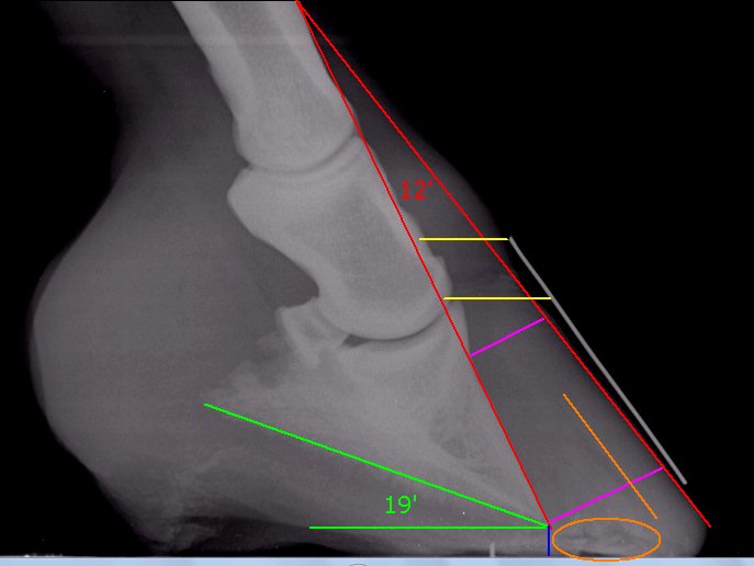
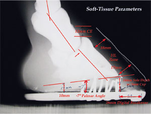
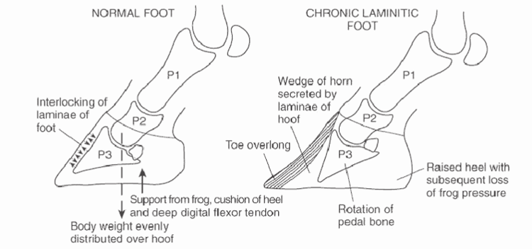
Laminitis radiograph measurements. Radiographic studies are an essential component in evaluation of horses with laminitis. Measurements can be made between the dorsal hoof wall and the dorsal aspect of the distal phalanx 1a and 1b. Lateromedial radiograph of a normal foot. Laminitis can affect one or all feet but it is most often seen in the front feet concurrently.
However founder usually refers to a chronic long term condition associated with rotation of the coffin bone whereas acute laminitis refers to symptoms associated with a sudden initial. The standard radiographs that should be obtained to aid assessment of horses with laminitis are the lateromedial horizontal dorsopalmar and dorsal 45 proximal palmarodistal oblique views. Laminitis laminitis survey horses radiography radiographic assessment radiography guidelines created date. Laminitis radiology chris pollitt the phalanges radiology of the equine limbs radiography john the vet.
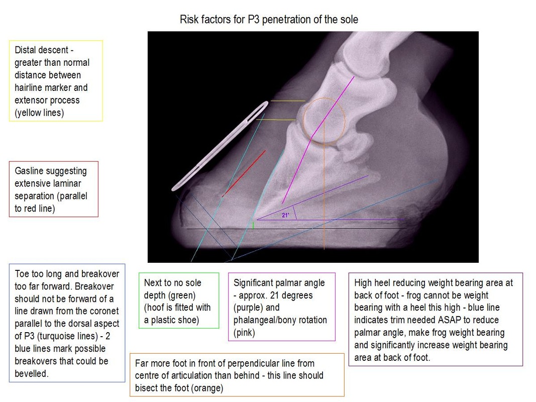
We can immediately see the additional information that can be gained from. Radiographic studies are an essential component in evaluation of horses with laminitis. Positioning techniques tips for acquiring good images shalyn j crawford 2015 textbook of veterinary diagnostic radiology donald e thrall 2013 p 245 247 explaining laminitis and its prevention robert eustace. Lateromedial radiographic projections were made from the feet of 25 normal horses and 3 angles and 3 distances were measured.
When us ing conventional and digital radiography techniques and views are well standardized 19 standard views including the lateral to medial dorsal palmar and 45 dorsal palmar projections should be performed rou tinely. It has evolved to where it quite beneficial for the farrier to use radiographs for guidance when trimming the equine foot. The terms laminitis and founder are used interchangeably. From these normal ranges of calculated variables were obtained.
Radiographic parameters measurement guide this page is meant to be used as an aid to measuring helpful parameters from a podiatry style farrier friendly radiographs. Strict guidelines with regards to radiographic geometry must be adhered too in order to obtain consistent findings between cases. X ray technique laminitis radiology chris pollitt the phalanges radiography john the vet. Radiology of the equine hoof is used to confirm various disease processes such as laminitis third phalanx fractures osteoarthritis ringbone navicular disease and extensive hoof wall separations.
Reading The Foot The Laminitis Site
The Laminitic Foundered Horse From The Acute To The Chronic Case
Laminitis And The Feet The Laminitis Site
Arming Yourself For The Battle With Laminitis American Farriers Journal
Understanding Laminitis Founder Southwest Equine Veterinary Group

Innovative Equine Podiatry Radiographic Parameters Measurement Guide

Innovative Equine Podiatry Acute Laminitis Case Study
Radiograph Of A Foot With Severe Chronic Laminitis Of 4 Weeks Duration Download Scientific Diagram

Palmar Angle Calculator The Laminitis Site

Laminitis A Pictorial Review

The Quest To Conquer Laminitis Horse Health Horses Types Of Bones
Laminitis Myths The Laminitis Site

Laminitis Or Founderthe Vet Centre Richmond

