Laminated Thrombus Ventricle

Thrombus An Overview Sciencedirect Topics

Thrombus An Overview Sciencedirect Topics

Rosh Review Aortic Aneurysm Abdominal Aortic Aneurysm Emergency Medicine
Example Of Thrombi As Depicted By Ice Panel A Small Apical Thrombus Download Scientific Diagram

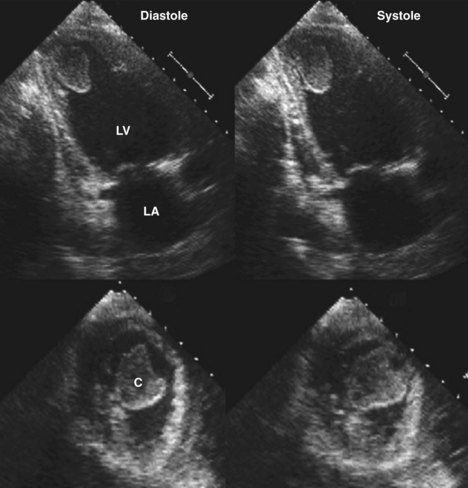
A B Left Ventricle Thrombi Are Shown 3 Chamber View In Two Different Download Scientific Diagram
Http Www Annalsthoracicsurgery Org Article S0003 4975 04 00209 7 Pdf

Typically the clot is a mural thrombus meaning it is on the wall of the ventricle.
Laminated thrombus ventricle. Of the 231 patients with echocardiographic evidence of thrombus resolution 20 8 7 developed a stroke or systemic embolism within 30 days after the echocardiogram. The next day she col lapsed suddenly with cyanosis tachypnea and hypotension with a slow idioventricular rhythm and died within a few minutes. Studies have demonstrated high incidence of lvt following anterior myocardial infarction lvt carries both short term and long term risk of embolic events which may result in stroke and systemic complications 2 3. Lvt is a common complication of acute myocardial infarction ami.
Left ventricular thrombus is a blood clot in the left ventricle of the heart. Department of internal medicine cardiovascular division university of texas southwestern medical center dallas texas usa summary. Intracardiac thrombi are seen in a variety of clinical settings and can result in severe morbidity or even death from embolic events they can occur following myocardial infarction with ventricular thrombus formation or with atrial fibrillation and mitral stenosis where atrial thrombi predominate. A type 1 excludes note is a pure excludes.
It means not coded here. Contemporary epidemiologic data suggest the incidence of lv thrombus detected using optimal imaging modalities may be as high as 15 in patients with st segment elevation mi stemi and up to 25 in patients with anterior mi. Left ventricular lv thrombus is a feared complication of lv dysfunction associated with high rates of systemic embolism morbidity and mortality. The primary risk of lvt is the occurrence of cardiac embolism in which the thrombus detaches from the ventricular wall and travels through the circulation and blocks.
Traditionally lv thrombus has been associated with acute myocardial infarction mi. At autopsy she was found to have laminated thrombus in the superior vena cava right atrium and ventricle but no evidence of pulmonary embolism. Kfeley m d and l. 19 83 86 1996 reviews left ventricular mural thrombus after acute myocardial infarction ellen c.
Lv thrombus is not an uncommon complication of acute mi and is associated with systemic thromboembolism. Left ventricular mural thrombus is a well recog nized complication of acute myocardial infarction. Left ventricular mural thrombus lvt complicating myocardial infarction has significant morbidity and potential mortality. Median follow up was 351 days interquartile range iqr 51 866 days.
Thrombi in the chambers of the left heart are a common source of complications like stroke and.

Now You See It Now You Don T Left Ventricular Thrombus Echopraxis
Echocardiographic Assessment Of Heart Failure Resulting From Coronary Artery Disease Thoracic Key

Heart Conditions Poster Heart Disease Anatomical Chart Company Heart Conditions Cardiology Medical

Heart Disease Poster Heart Pathology Anatomical Chart Company Heart Anatomy Congenital Heart Disease Cardiology

Ryan Here S The Basics Hope This Helps Nurse Medical Nursing Information

Max Brodel Heart Amazing Art Art Artsy Fartsy

Deep Vein Thrombosis Chart 20x26 Vein Thrombosis Deep Vein Thrombosis Thrombosis

Signs And Symptoms Of Pulmonary Embolism Calgary Guide Pulmonary Embolism Pulmonary Embolism Nursing Pulmonary

Rosh Review Medical Anatomy Icu Nursing Medical School Studying

The Left Panel Shows An Echogenicity At The Apex Suspicious Of A Left Download Scientific Diagram

Extensive Pulmonary Embolism Pulmonary Embolism Pulmonary Pulmonary Embolism Nursing

The Human Brain Laminated Anatomy Chart Human Brain Anatomy Human Brain Brain Anatomy

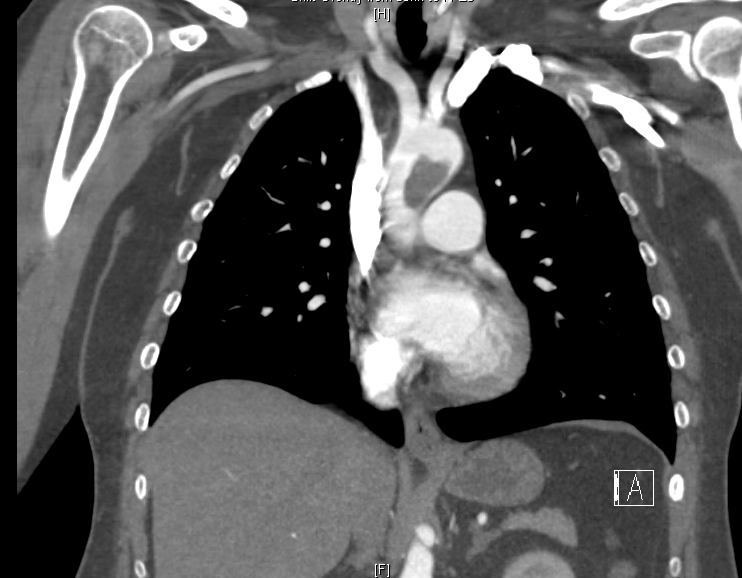
Ct Angiography Showing A Thrombus In The Pulmonary Artery Branches Download Scientific Diagram