Laminated Lv Apical Thrombus
Echocardiographic Assessment Of Heart Failure Resulting From Coronary Artery Disease Clinical Gate
Http Www Annalsthoracicsurgery Org Article S0003 4975 04 00209 7 Pdf

Thrombus An Overview Sciencedirect Topics

How To Assess A Left Ventricular Thrombus With The Help Of Cardiac Mri Cmr Youtube

The Left Panel Shows An Echogenicity At The Apex Suspicious Of A Left Download Scientific Diagram
Example Of Thrombi As Depicted By Ice Panel A Small Apical Thrombus Download Scientific Diagram

Follow up occurred through march 2019.
Laminated lv apical thrombus. Mural thrombi are most commonly seen between six and 10 days following an acute myocardial infarction mi. Before cardioversion 17 81 patients were anticoagulated with warfarin or heparin. Patients with apical myocardial infarction are at higher risk of developing left ventricular lv thrombi. B shows the thrombus in the left atrium la before removal of the.
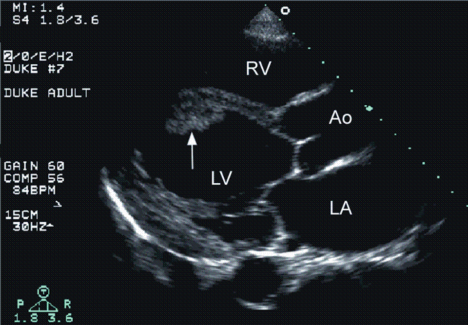
Standard transesophageal echocardiography tee is commonly used in assessing cardiac source of embolic cerebrovascular accident cva. Left ventricular mural thrombus lvt complicating myocardial infarction has significant morbidity and potential mortality. A represents the precardiopulmonary bypass image of the apical thrombus in the left ventricle lv. Typically the clot is a mural thrombus meaning it is on the wall of the ventricle.
Left ventricular lv thrombosis persists as a clinical challenge in echocardiographic diagnosis and is an important risk factor for perioperative embolic events in cardiac surgery. Left ventricular lv thrombus is a feared complication of lv dysfunction associated with high rates of systemic embolism morbidity and mortality. Lvt is a common complication of acute myocardial infarction ami. Appropriate detection and monitoring when thrombus is suspected is critical in surgical planning and in avoiding catastrophic patient outcomes.
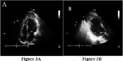
Lvt carries both short term and long term risk of embolic events which may result in stroke and systemic complications 2 3. Studies have demonstrated high incidence of lvt following anterior myocardial infarction. A three center cohort study was performed identifying 514 patients with lv thrombus on echocardiography between october 2013 and march 2019. Transesophageal echocardiography images of case 2.
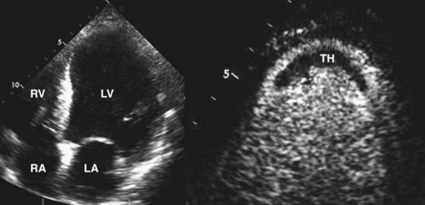
Traditionally lv thrombus has been associated with acute myocardial infarction mi. They occur at the left ventricular apex and are more common following an anterior wall infarction since anterior mis involve more of the apex fig 2.

Figure 1 From Leaking Left Ventricular Pseudoaneurysm Semantic Scholar
Intracardiac Masses Springerlink

Now You See It Now You Don T Left Ventricular Thrombus Echopraxis

A B Left Ventricle Thrombi Are Shown 3 Chamber View In Two Different Download Scientific Diagram

Long Term Consequences And Prognosis After Myocardial Infarction Thoracic Key
Https Www Ahajournals Org Doi Pdf 10 1161 01 Cir 58 3 528
Https Www Onlinejase Com Article S0894 7317 05 00985 5 Pdf

Ventricular Tachycardia Ablation In The Presence Of Left Ventricular Thrombus Safety And Efficacy Rao 2016 Journal Of Cardiovascular Electrophysiology Wiley Online Library
2
Https Journals Sagepub Com Doi Pdf 10 1177 87579302018003001

Pdf The Use Of Echocardiography In Predicting Left Ventricle Thrombus In Patients With Idiopathic Dilated Cardiomyopathy At Chris Hani Baragwanath Hospital Semantic Scholar

Echocardiographic Assessment Of Heart Failure Resulting From Coronary Artery Disease Thoracic Key
Adrenal Insufficiency Induced Cardiomyopathy Clinical Cardiology And Cardiovascular Medicine Addisons Disease

