Laminated Left Ventricular Thrombus

The Left Panel Shows An Echogenicity At The Apex Suspicious Of A Left Download Scientific Diagram
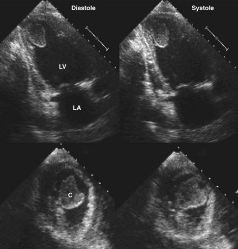
Echocardiographic Assessment Of Heart Failure Resulting From Coronary Artery Disease Thoracic Key

How To Assess A Left Ventricular Thrombus With The Help Of Cardiac Mri Cmr Youtube
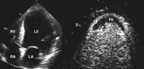
Echocardiographic Assessment Of Heart Failure Resulting From Coronary Artery Disease Clinical Gate
Http Www Annalsthoracicsurgery Org Article S0003 4975 04 00209 7 Pdf

A B Left Ventricle Thrombi Are Shown 3 Chamber View In Two Different Download Scientific Diagram
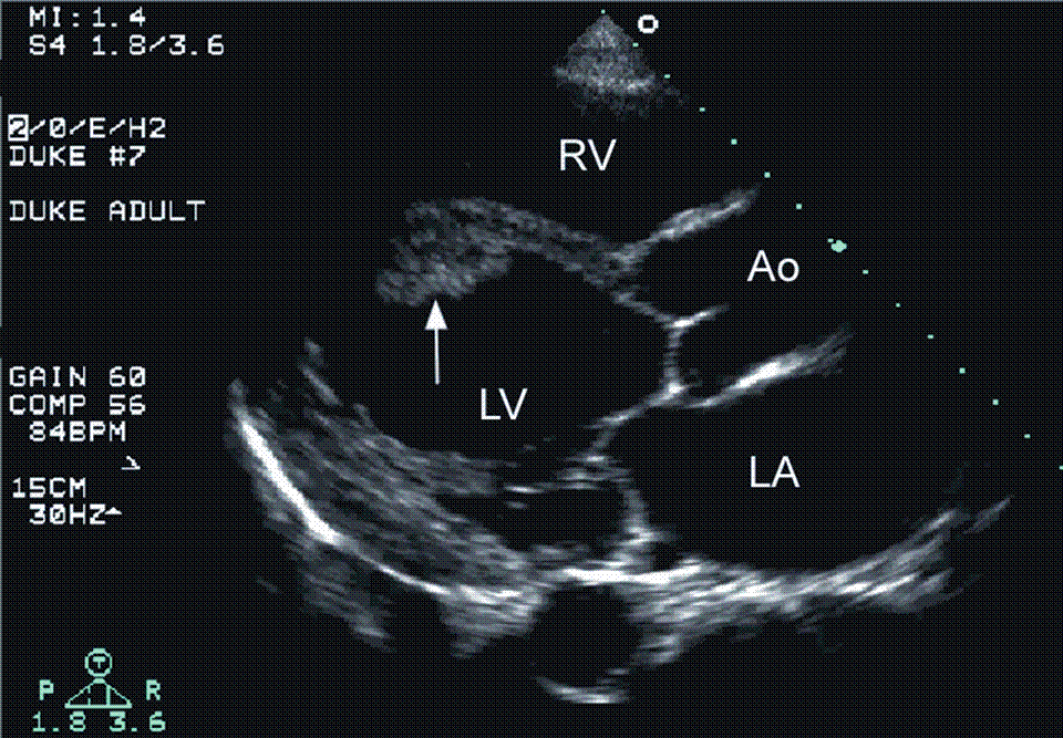
Identification of left ventricular mural thrombus lvt may be challenging depending on the imaging modality used.
Laminated left ventricular thrombus. Left ventricular thrombus lvt complicates both ischemic and non ischemic cardiomyopathies and is a potential cause of thromboembolic complications such as stroke. Acute procedural outcomes complications and clinical outcomes at 1 year were assessed. Appropriate detection and monitoring when thrombus is suspected is critical in surgical planning and in avoiding catastrophic patient outcomes. Typically the clot is a mural thrombus meaning it is on the wall of the ventricle.
Left ventricular thrombus is a blood clot thrombus in the left ventricle of the heart. Left ventricular thrombus formation after myocardial infarction. Traditionally lv thrombus has been associated with acute myocardial infarction mi. A calcified lvt was defined as a persistent left ventricular mural thrombus encapsulated by thickened and calcified endocardium.
Left ventricular characteristics including left ventricular ejection fraction lvef left ventricular volume wall motion cardiac output and potential mechanical complications were also collected. A sixty four years old female presented with worsening dyspnea on exertion with troponin elevation. Lvt is a common complication of acute myocardial infarction ami. Left ventricular lv mural thrombus is a well recognized complication of acute myocardial infarction.
Patients with laminated lv thrombus on transthoracic echocardiogram who underwent scar mediated vt ablation at two centers from 2010 to 2013 were retrospectively analyzed. All patients had failed medical therapy. Left ventricular lv thrombus is a feared complication of lv dysfunction associated with high rates of systemic embolism morbidity and mortality. We present a case of lvt which was incidentally identified on cine cardiac magnetic resonance imaging cmr.

Thrombus An Overview Sciencedirect Topics

Long Term Consequences And Prognosis After Myocardial Infarction Thoracic Key

Now You See It Now You Don T Left Ventricular Thrombus Echopraxis

Figure 1 From Leaking Left Ventricular Pseudoaneurysm Semantic Scholar

Ventricular Tachycardia Ablation In The Presence Of Left Ventricular Thrombus Safety And Efficacy Rao 2016 Journal Of Cardiovascular Electrophysiology Wiley Online Library
Https Www Ahajournals Org Doi Pdf 10 1161 01 Cir 58 3 528
2
Intracardiac Masses Springerlink

Thrombus An Overview Sciencedirect Topics
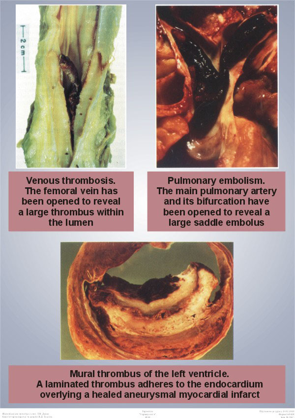
Venous Thrombosis Pulmonary Embolism Mural Thrombus Of The Left Ventricle

Hma Lectures Thrombosis Studyingmed

Medmastery How To Assess Lv Thrombus Facebook
Https Onlinelibrary Wiley Com Doi Pdf 10 1111 Echo 13431

