Laminated Apical Thrombus
Echocardiographic Assessment Of Heart Failure Resulting From Coronary Artery Disease Thoracic Key
Example Of Thrombi As Depicted By Ice Panel A Small Apical Thrombus Download Scientific Diagram
Http Www Annalsthoracicsurgery Org Article S0003 4975 04 00209 7 Pdf

How To Assess A Left Ventricular Thrombus With The Help Of Cardiac Mri Cmr Youtube

Thrombus An Overview Sciencedirect Topics

Now You See It Now You Don T Left Ventricular Thrombus Echopraxis

Mural thrombus is basically a blood clot that is formed in the blood and is attached to the lining of a chamber of the heart or the wall of a blood vessel.
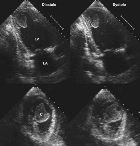
Laminated apical thrombus. This thrombus is seen in both a parasternal long axis view left and an apical four chamber view right. Farlex partner medical dictionary farlex 2012 want to thank tfd for its existence. They are dangerous and can break loose to form emboli. Contemporary epidemiologic data suggest the incidence of lv thrombus detected using optimal imaging modalities may be as high as 15 in patients with st segment elevation mi stemi and up to 25 in patients with anterior mi.
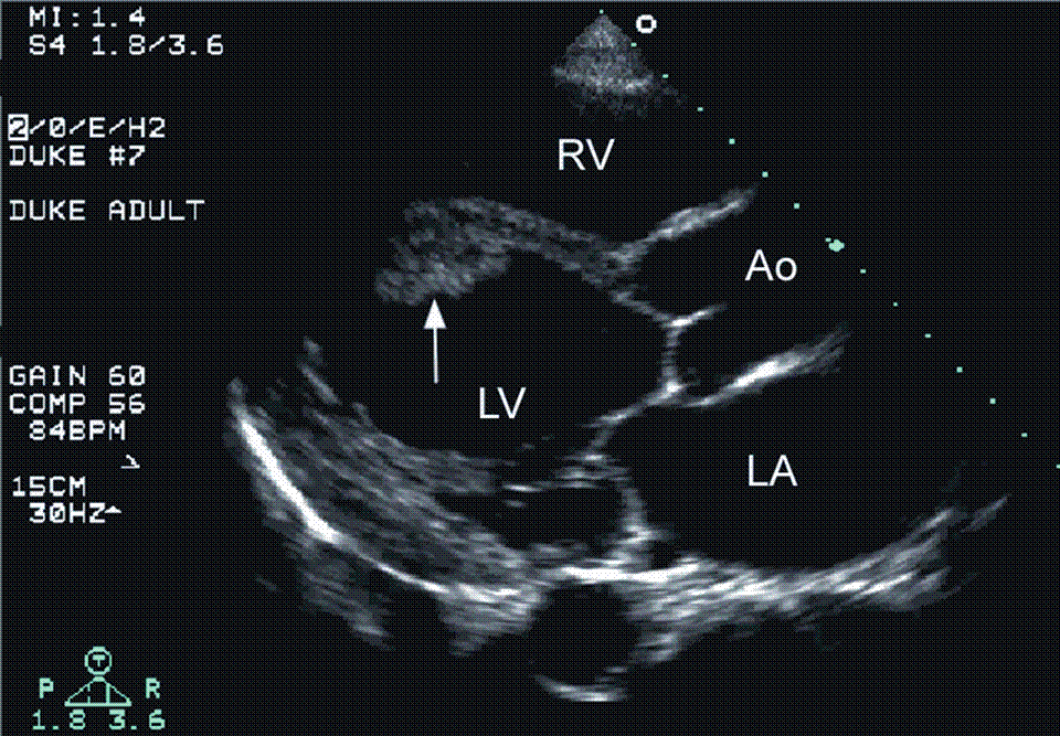
Lack of echo contrast uptake by a laminated thrombus in an aneurysmal basal inferior wall camouflaged as the myocardium itself and hence likely concealed the thrombus. The authors present a case of a laminated lv apical thrombus which was discovered intraoperatively by real time 3 dimensional 3d transesophageal echocardiography. Lv thrombus is not an uncommon complication of acute mi and is associated with systemic thromboembolism. Lam i nat ed throm bus a thrombus formed gradually by clotting of the blood in successive layers.

Figure 1 From Leaking Left Ventricular Pseudoaneurysm Semantic Scholar

The Left Panel Shows An Echogenicity At The Apex Suspicious Of A Left Download Scientific Diagram

A B Left Ventricle Thrombi Are Shown 3 Chamber View In Two Different Download Scientific Diagram
Adrenal Insufficiency Induced Cardiomyopathy Clinical Cardiology And Cardiovascular Medicine Addisons Disease
Https Www Ahajournals Org Doi Pdf 10 1161 01 Cir 58 3 528
Https Www Onlinejase Com Article S0894 7317 05 00985 5 Pdf

Pdf The Use Of Echocardiography In Predicting Left Ventricle Thrombus In Patients With Idiopathic Dilated Cardiomyopathy At Chris Hani Baragwanath Hospital Semantic Scholar
Intracardiac Masses Springerlink

The Dancing Thrombus 4d Transesophageal Echocardiography In The Diagnosis Of A Pedunculated Left Atrial Appendage Thrombus Kelly 2018 Echocardiography Wiley Online Library

Long Term Consequences And Prognosis After Myocardial Infarction Thoracic Key

Pin On Medicine

