Lamin A C Structure
Schematic Presentation Of Lamin A C And The Hbcag Ig Fold Fusion Download Scientific Diagram

Structure Of The Asymmetric Unit Of The Human Lamin A C Fragment A Download Scientific Diagram
The Umd Lmna Database The Protein

Images Cell Organelles Organelles Functions Organelles

Pin On Science

Comparison Of Lamin A Lamin C And La 50 Structures A The 12 Download Scientific Diagram

Lamin a c antibody e 1 is available as both the non conjugated anti lamin a c antibody form as well as multiple conjugated forms of anti lamin a c antibody including agarose hrp pe fitc and multiple alexa fluor conjugates.
Lamin a c structure. During apoptosis lamin a c is specifically cleaved into a large 41 50 kda and a small 28 kda fragment 3 4. A unique family of cysteine proteases has been described that differs in sequence structure and substrate. The lamin family of proteins make up the matrix and are highly conserved in evolution. 2000 j struct biol 129 313 23.
Lamins are grouped into a type and b type lamins. Lamin a c is cleaved by caspase 6 and serves as a marker for caspase 6 activation. 1 product result match criteria. Lamin proteins are thought to be involved in nuclear stability chromatin structure and gene expression.
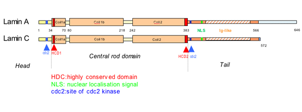
1989 showed that lamin a is synthesized as a precursor molecule called prelamin a. Lamin a c is an intermediate filament lining the inner nuclear membrane part of the nuclear envelope. In mammals alternative splicing gives rise to lamin a and lamin c and the less abundant isoforms lamin aδ10 and lamin c2 fisher et al 1986. A type lamins are encoded by a single gene lmna.
290 the transcript from the gene is spliced differentially to give rise to the two different forms lamin a and lamin c. Maturation of lamin a involves the removal of 18 residues from the c terminus which is accomplished by isoprenylation and farnesylation involving a c terminal caax cysteine aliphatic aliphatic any amino acid box sinensky et al 1994. Lamin proteins are thought to be involved in nuclear stability chromatin structure and gene expression. The lack of lamin a c can be as a novel marker for undifferentiated embryonic stem cells and lamin a c expression is as an early indicator of differentiation pmid.
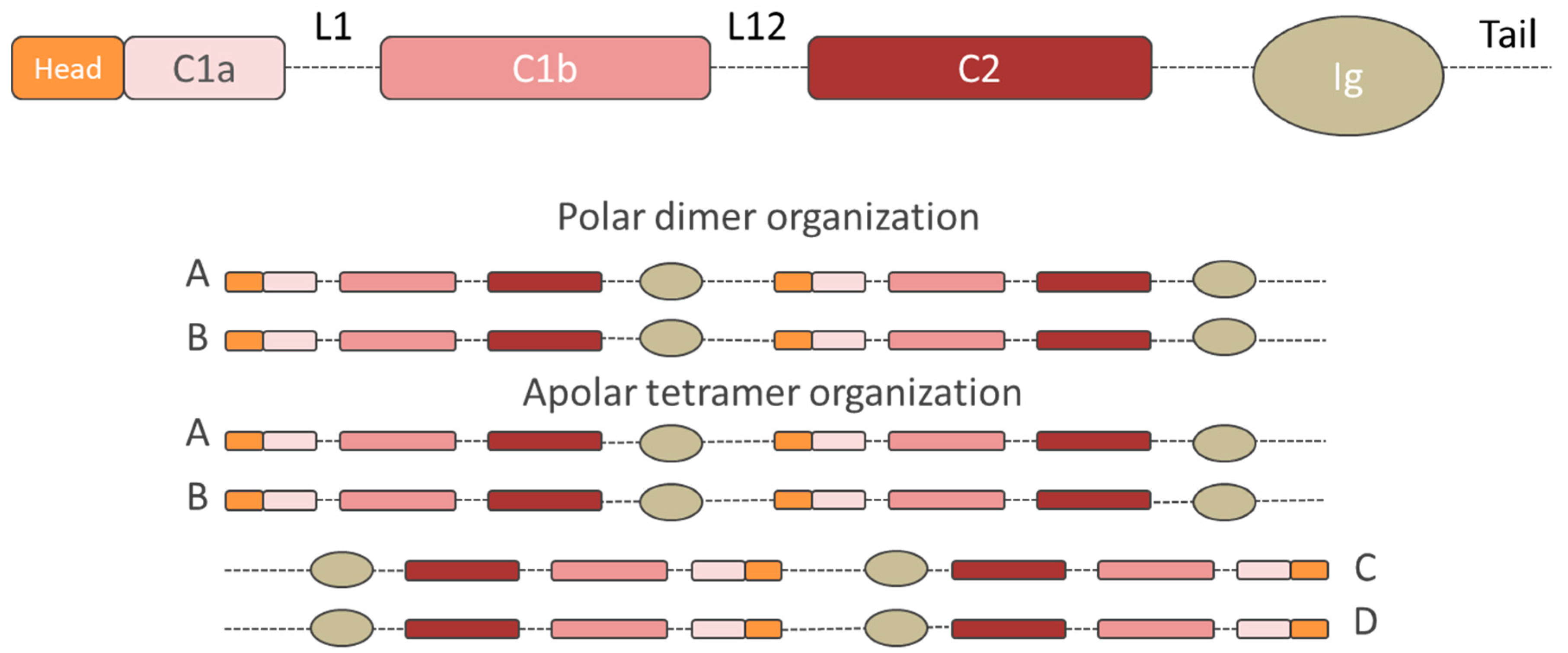
These molecules bind to other inner nuclear membrane proteins such as emerin which is mutated in x linked edmd but. During mitosis the lamina matrix is reversibly disassembled as the lamin proteins are phosphorylated. Mutations in the region of human lmna encoding the carboxyl terminal tail lamin a c are associated with forms of muscular dystrophy and familial partial lipodystrophy fpld. Lamin structure and assembly.
Note the presence of irregularly shaped nuclear envelopes in many of the subject s fibroblasts lamins also known as nuclear lamins are fibrous proteins in type v intermediate filaments providing structural function and transcriptional regulation in the cell nucleus. Vertebrate lamins consist of two types a and b. Furukawa et al 1994. Anti lamin a c antibody clone 2e8 2 antibody alexa fluor 488 conjugate.
The cleavage of lamins results in nuclear dysregulation and cell death 5 6.
Cells Free Full Text Lamin A C Mechanotransduction In Laminopathies

What A Blob Chromosomes Rarely X Shaped Chromosome Structure Chromosome Medical Art

Structure Of Lamin A Protein A Diagram Of Lamin A Amino Acid Sequence Download Scientific Diagram

Figure 1 From The Nuclear Lamins Flexibility In Function Semantic Scholar

Structural Organization Of Nuclear Lamins A C B1 And B2 Revealed By Superresolution Microscopy Molecular Biology Of The Cell

When Lamins Go Bad Nuclear Structure And Disease Cell

Broken Nuclei Lamins Nuclear Mechanics And Disease Sciencedirect

Lamin Structure A Schematic Sketch Of A Generic Lamin Protein Download Scientific Diagram

Assessment Of Genetic Mutations Dmd Dysf Emd Lmna Dux4 Dmpk Znf9 Pabpn1 Genes Induction Du Life Science Duchenne Muscular Dystrophy Muscular Dystrophies

Structure Of The Nucleus Labeled Cell Organelles Organelles Cell Model

Assembly Properties Of Gfp Tagged Lamin Constructs A Domain Download Scientific Diagram

Lmna An Overview Sciencedirect Topics
Rcsb Pdb 6jlb Crystal Structure Of Lamin A C Fragment And Assembly Mechanisms Of Intermediate Filaments

