Lamin A C Staining

Immunofluorescence Staining Of Lamins A And C In Cells From Metastatic Download Scientific Diagram

Lamin A C Antibody Nbp2 25152 Novus Biologicals

Lamin A C Staining Of Muscles Sections Of Rag Mdx Muscles Transplanted Download Scientific Diagram
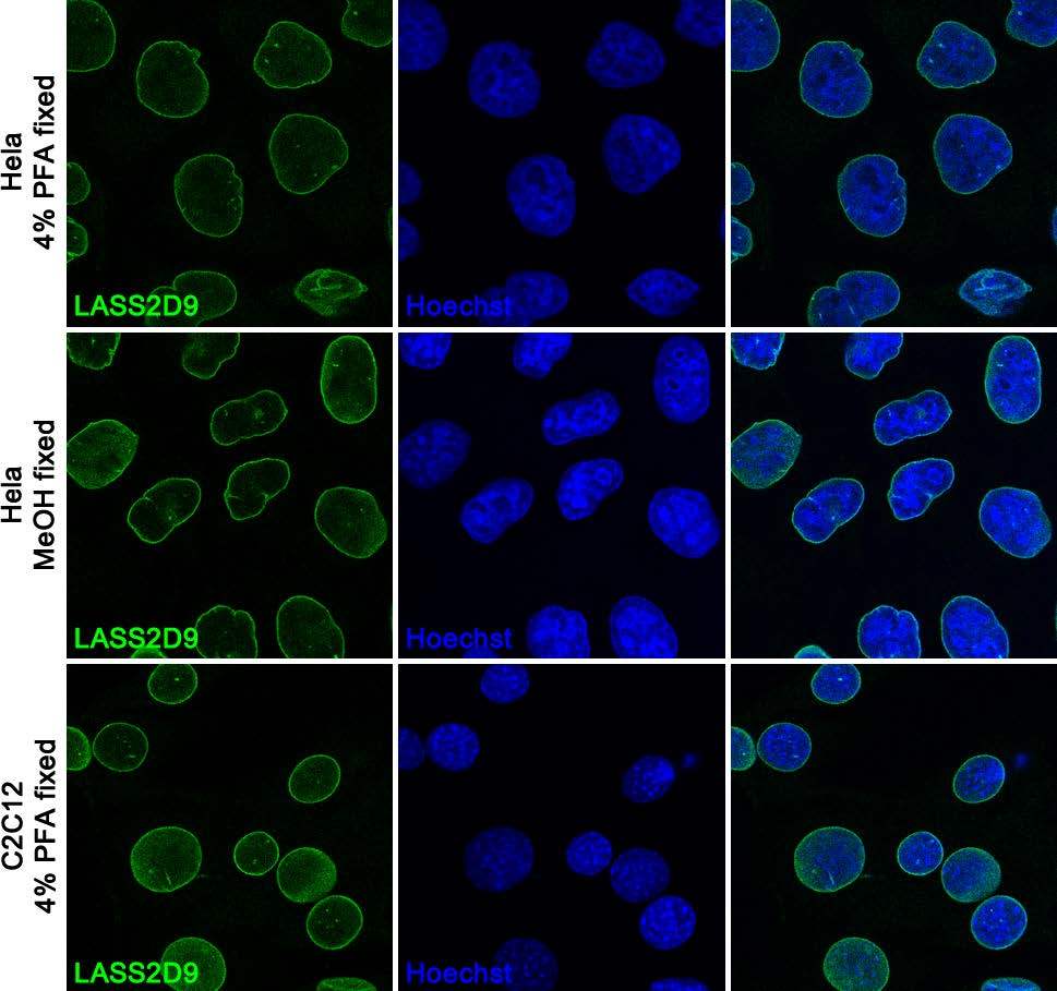
Anti Lamin A C Lass2d9 Monoclonal Antibodies Ximbio

Nuclear Lamins Localization At The Nuclear Periphery And Within The Download Scientific Diagram

7497 Phospho Ndrg1 Thr346 D98g11 Xp Rabbit Mab Alexa Fluor 647 Conjugate Cst抗体 Confocal Immunofluorescent Analysis Of Mab Painting Alexa
Farnesylation of prelamin a c facilitates nuclear envelope targeting and subsequent cleaveage by zmpste24 face1 to remove the farnesyl group produces mature lamin a c which can then be inserted into the nuclear lamina.
Lamin a c staining. Lamin a c is cleaved by caspase 6 and serves as a marker for caspase 6 activation. I reviewed the provided product datasheet and this rabbit anti mouse antibody would fall into this category and should for this reason detect the ab8984. Many immature type 2c intermediate staining muscle fibers. The lamin a c lmna gene contains 12 exons.
Fiber type changes type 1 predominance atpase ph 9 4 stain. Lamin a c antibody nb100 56649 intracellular staining of hela cells 1 x 10 6 cells ml with lamin a c antibody orange stained at 1 1000. Congenital muscular dystrophy lamin a c. Lamin a c green f actin staining with phalloidin red and nuclei with dapi blue is shown.
There are three types of lamins a b and c. I can confirm that any anti mouse antibody that detects the complete igg isotype or the igg1 isotype should detect the anti lamin a c antibody 131c3 nuclear envelope marker ab8984. Cells were probed without control or with an antibody recognizing lamin a c product ma3 1000 at a dilution of 1 20 over night at 4 c washed with pbs and. The anti lamin a c antibody reveals strong nuclear lamina staining while anti lamp1 antibody reveals strong cytoplasmic punctate staining of lysosomes and early endosomes.
During apoptosis lamin a c is specifically cleaved into a large 41 50 kda and a small 28 kda fragment 3 4. 2000 j struct biol 129 313 23. Since both dna blue and lamin a c red are associated with the nuclear compartment this region appears crimson in this image. Lamin a c antibody staining protocol for immunohistochemistry.
Cells were grown on chamber slides and fixed with formaldehyde prior to staining. Detected with a gtxrb dylight 488 secondary.

Lamin A C Antibody Phosphosolutions

What Is One Step Immunostaining
Cells Topical Collection Lamins And Laminopathies
-Immunocytochemistry-Immunofluorescence-NB100-74451-img0011.jpg)
Lamin A C Antibody Mab636 Nb100 74451 Novus Biologicals

Lamin A C Controls Nuclear Matrin 3 Levels And Localization But Not Alternative Splicing Of Cassette Exons Biorxiv
Lamin A C And Emerin Depletion Impacts Chromatin Organization And Dynamics In The Interphase Nucleus Springerlink

A Progeria Mutation Reveals Functions For Lamin A In Nuclear Assembly Architecture And Chromosome Organization Pnas
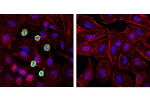
Phospho Lamin A C Ser22 D2b2e Xp Rabbit Mab Cell Signaling Technology

Figure 3 From Interphase Phosphorylation Of Lamin A Semantic Scholar

Confocal Image Of Some Dividing Cells That Are Triple Labeled For Golgi Apparatus Green Microtubules Red Science And Nature Mitosis Cell Biology
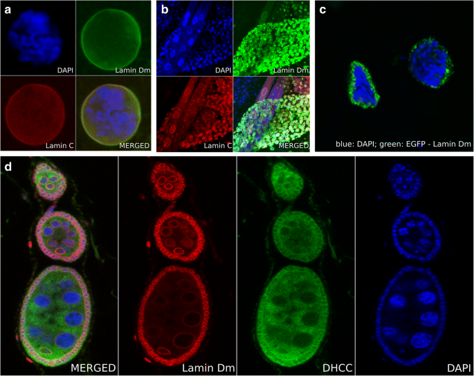
Laminopathies What Can Humans Learn From Fruit Flies Springerlink

Iridescent Blue Tile Blue Tiles My Favorite Color Dream Furniture

Lamin A C Maintains Exocrine Pancreas Homeostasis By Regulating Stability Of Rb And Activity Of E2f Gastroenterology
